Hemivertebra is a congenital spinal deformity in which one side of a vertebra fails to develop completely during fetal development. As a result, the affected vertebra forms a wedge-shaped bone rather than a normal rectangular structure. This asymmetry can lead to abnormal curvature of the spine.
Hemivertebra can occur anywhere along the spine, including the cervical, thoracic, or lumbar regions, and is commonly associated with spinal deformities such as scoliosis or kyphosis.
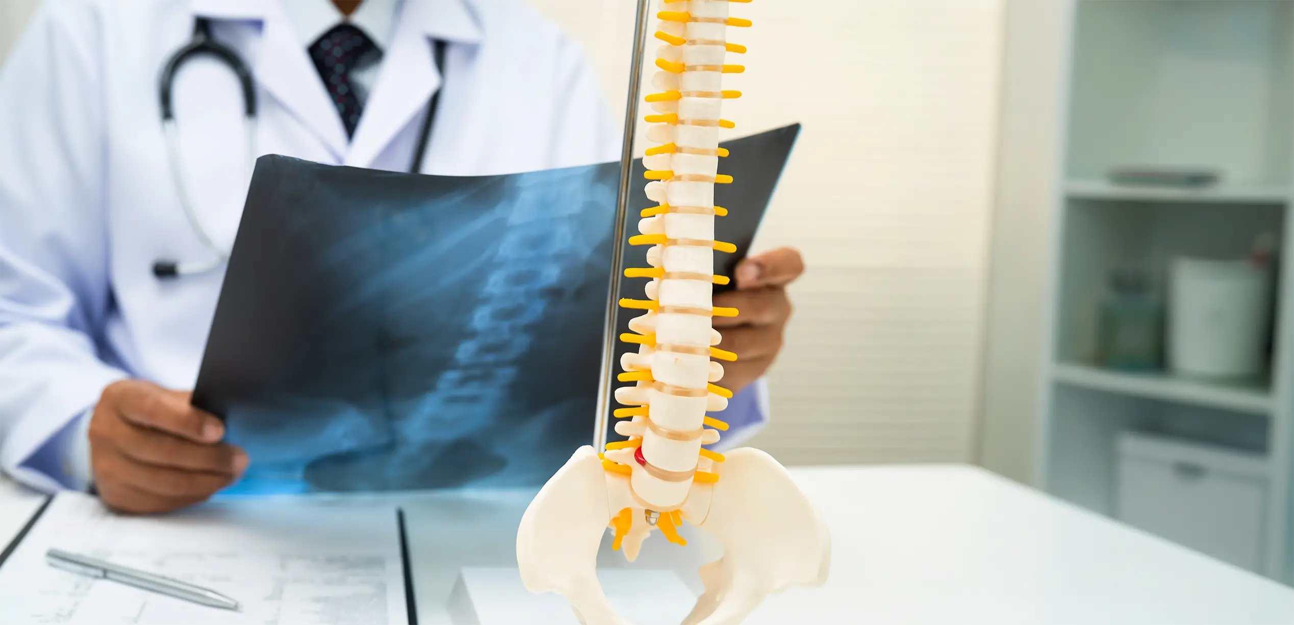
At Fakeeh University Hospital Dubai, our spine consultants provide comprehensive evaluation and individualized treatment for patients with congenital spinal deformities such as hemivertebra. Using advanced imaging and modern surgical techniques when required, our goal is to correct spinal alignment, maintain neurological safety, and support healthy spinal development.
Early diagnosis and proper management are essential to prevent progression of spinal deformity and long-term complications.
The spine normally develops from symmetrical vertebral segments during early fetal growth. In hemivertebra, only one half of a vertebra forms properly, while the other half remains underdeveloped or absent.
This structural imbalance can cause the spine to curve as the child grows. Depending on the location and severity, the deformity may progress over time.
Some cases remain mild, while others may lead to significant spinal curvature.
Hemivertebra develops during embryonic spinal formation, when a vertebra fails to develop completely.
Although the exact cause is not always known, several factors may contribute:
In many cases, hemivertebra occurs as an isolated spinal condition, but it may sometimes be associated with other congenital abnormalities.
Different forms of hemivertebra may occur depending on how the vertebra develops.
This is the most common form. The incomplete vertebra develops on one side, leading to lateral curvature of the spine, commonly resulting in scoliosis.
In this type, the defect affects the posterior portion of the vertebra, which may contribute to excessive forward curvature of the spine known as kyphosis.
In complete hemivertebra, half of the vertebra is entirely absent, which may lead to more pronounced spinal deformity.
In some cases, hemivertebra may not cause symptoms initially, particularly in mild deformities. However, as the spine grows, symptoms may develop.
Common symptoms include:
In more severe cases, spinal deformity may lead to compression of the spinal cord or nerves, which can cause:
These symptoms require urgent medical evaluation.
At Fakeeh University Hospital Dubai, diagnosis begins with a detailed clinical assessment.
The physician evaluates spinal alignment, posture, and any visible curvature.
Assessment of neurological function is also important to determine whether the spinal cord or nerves are affected.
Imaging tests are essential to confirm the diagnosis and evaluate the extent of the deformity.
These may include:
These studies help guide appropriate treatment planning.
Treatment depends on several factors, including the severity of the spinal deformity, patient age, and the progression of the curvature.
In mild cases, particularly in young children, careful monitoring with periodic imaging may be recommended to observe spinal growth and detect any progression of deformity.
For moderate spinal curvature, spinal bracing may help stabilize the spine and limit progression while the child grows.
Surgery may be recommended when:
Surgical treatment may involve removal of the hemivertebra and stabilization of the spine using rods and screws.
The goal of surgery is to correct spinal alignment, prevent further curvature progression, and maintain neurological safety.
Modern surgical techniques allow precise correction while preserving spinal stability.
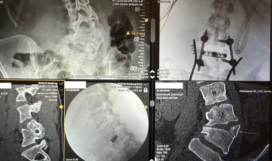
A 20-year-old woman from Germany presented with severe back and leg pain caused by a neglected L4 hemivertebra, which had produced a left-convex lumbar scoliosis and compression of the right nerve roots. Using a minimally invasive approach, we decompressed the affected nerves, resected the hemivertebra, and stabilized the spine. The deformity was corrected with only a single-segment fusion, following the principle “as much as necessary, as little as possible.” This allowed preservation of spinal mobility in a young patient. Back and leg pain resolved completely, and the spinal curve was corrected to a great extent.
With early detection and appropriate treatment, many patients with hemivertebra achieve good outcomes and maintain normal spinal function.
Regular monitoring during growth is important to detect changes early and guide appropriate intervention.
Medical evaluation is recommended if you notice:
Early specialist evaluation helps ensure the best possible treatment outcome.
If you suspect spinal deformity such as hemivertebra or notice abnormal spinal curvature, early evaluation is important.
Book your appointment at Fakeeh University Hospital Dubai to receive expert diagnosis and specialized spine care.

Hemivertebrae is a congenital spinal condition in which one or more vertebrae develop incompletely — forming a wedge-shaped half vertebra instead of a normal, symmetrical one. This abnormal vertebral shape disrupts the natural alignment of the spine, often leading to scoliosis (sideways curvature) or kyphosis (forward curvature).
The exact cause of hemivertebrae is not always known, but contributing factors may include:
Hemivertebrae may occur as a single isolated defect or alongside other congenital abnormalities affecting the heart, kidneys, or limbs.
Hemivertebrae in newborns may be suspected at birth if visible spinal curvature, asymmetry of the back, or other associated anomalies are noted during the neonatal physical examination.
Diagnostic steps typically include:
Early and accurate diagnosis in newborns is critical for monitoring spinal progression and planning appropriate treatment before significant curvature develops.
On radiological imaging, hemivertebrae has characteristic appearances that help specialists make an accurate diagnosis:
On X-ray:
On CT scan:
On MRI:
Radiological assessment by a specialist musculoskeletal radiologist is essential for accurate classification and treatment planning.
Yes — hemivertebrae can sometimes be detected on fetal ultrasound during pregnancy, though detection depends on the position of the baby, the quality of imaging, and the severity of the defect.
Key points about hemivertebrae fetal ultrasound:
If hemivertebrae is suspected on fetal ultrasound, referral to a specialist in fetal medicine and a paediatric spine surgeon is recommended for early planning and counselling.


.webp)