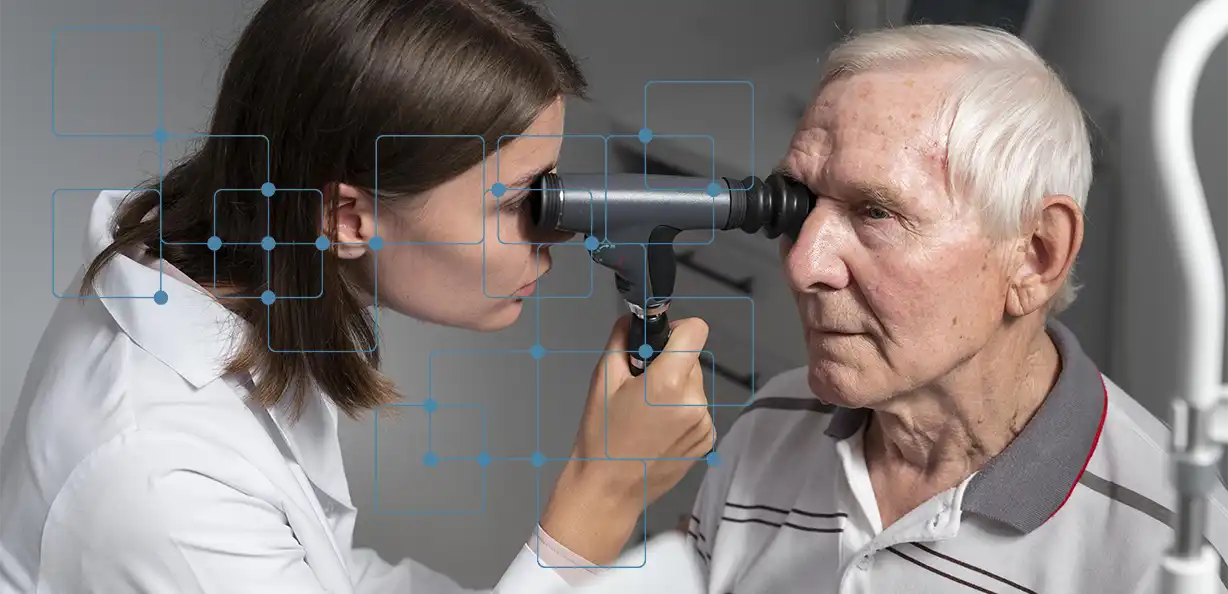
A general physician may refer a patient to an ophthalmologist if he or she shows symptoms of eye infections, optic nerve problems, cataracts, or other eye problems. Our Ophthalmic Department at Fakeeh University Hospital delivers quality eye care in an ethical and humanistic manner.
Our ophthalmologists are trained and qualified to use the latest technology to offer quality medical eye care. They can diagnose, prevent, and treat of almost all eye conditions and visual problems.
In addition to caring for the eyes and vision, our ophthalmologists have undergone medical training that enables them to notice symptoms of conditions that do not directly relate to the eye. In such instances, they can refer people for appropriate treatment to the right departments. We have best eye doctor for glaucoma. They also take part in some scientific research that focuses on the causes of eye and vision conditions, and potential treatments.


Treatment of Cornea and Ocular Surface Disease:

We have a specialized glaucoma unit with different treatment modalities. You will get a complete eye exam, effective solutions for your vision needs.
The Outpatient Clinic utilizes cutting-edge diagnostic equipment, including:
Clinical Applications
If you are searching for an eye specialist near you or an optometrist near you, look no further than Fakeeh University Hospital in Dubai. We provide a range of eye care services, from eye exams near you and vision tests near you to comprehensive check-ups and treatment for various eye conditions. Our team includes some of the best professionals, such as the best optometrist Dubai and ophthalmologist Dubai near you, who are ready to help you maintain optimal vision health. Whether you need medical eyeglasses or are looking for the best eye doctor for glaucoma, we are here to cater to your needs. Schedule an eye appointment near you with us at the nearest eye hospital in Silicon Oasis Dubai, and experience exceptional care with state-of-the-art facilities. For an eye check up near you or just needing routine eye check up, visit Fakeeh University Hospital Ophthalmology department to meet the specialised eye doctor and ensure your eyes stay healthy.
Clinical Applications
Clinical Applications
Clinical Applications
For the treatment of:
For the treatment of:

Common eye diseases that can cause permanent damage include glaucoma, macular degeneration, diabetic retinopathy, and advanced cataracts. Early detection and treatment can help prevent vision loss.

Glaucoma cannot be cured, but it can be managed effectively with medications, laser treatments, or surgery to prevent further vision loss. Early diagnosis is critical for successful management.

Non-serious causes of eye pain include dry eyes, eye strain, allergies, and mild infections like conjunctivitis. These can often be treated with lubricating drops or over-the-counter remedies.

During your first eye consultation, the doctor will review your medical history, conduct a comprehensive eye exam, and perform tests like vision acuity and eye pressure measurements. They may recommend further tests if needed.

A routine eye checkup includes assessing visual acuity, testing for common eye diseases like glaucoma or cataracts, checking eye pressure, and examining the health of your retina and optic nerve.

